What Does Cancer Look Like On Mammogram - Mammography Cuts Risk For Fatal Breast Cancers New Data / The look of breast cancer on a mammogram a tumor or lump will appear as a focused white area on the mammogram.. The tumor cells don't stay within the clear borders of the mass, but instead invade the nearby breast tissue. What does breast cancer look like on a digital mammogram. It's usually firm and rubbery, and doesn't hurt. Calcifications are calcium deposits within the breast tissue and they look like small white spots. They will look carefully at the mammogram to interpret the results.
What does breast cancer look like? Breast cancer and some noncancerous (benign) breast conditions can appear white on a mammogram. Cancers may be seen as masses (like a ball, but usually with an irregular shape), areas of asymmetry that resemble normal tissue, calcifications (white specks), and/or areas of architectural distortion (imagine the puckering caused by pulling a thread in a piece of fabric). Abnormalities such as cancerous tumors usually appear brighter because they are denser. What does breast cancer look like on a mammogram?
3d Mammograms Increasing In Popularity For Breast Cancer Screening from www.news-medical.net This is considered an abnormal mammogram, but not necessarily one that's indicative of cancer. A fibroadenoma can look like a small marble, and you can move it under your skin. Cancers may be seen as masses (like a ball, but usually with an irregular shape), areas of asymmetry that resemble normal tissue, calcifications (white specks), and/or areas of architectural distortion (imagine the puckering caused by pulling a thread in a piece of fabric). However, cancer can also be seen as white, making it a bit obscure in women with extremely dense breasts. Calcifications are tiny flecks of calcium — like grains of salt — in the soft tissue of the breast that can sometimes indicate the presence of an early breast cancer. The mammogram and all other tests came back normal. There are few risks associated with mammography. Invasive breast cancer can appear as a white patch or mass on a mammogram.
Cancers may be seen as masses (like a ball, but usually with an irregular shape), areas of asymmetry that resemble normal tissue, calcifications (white specks), and/or areas of architectural distortion (imagine the puckering caused by pulling a thread in a piece of fabric).
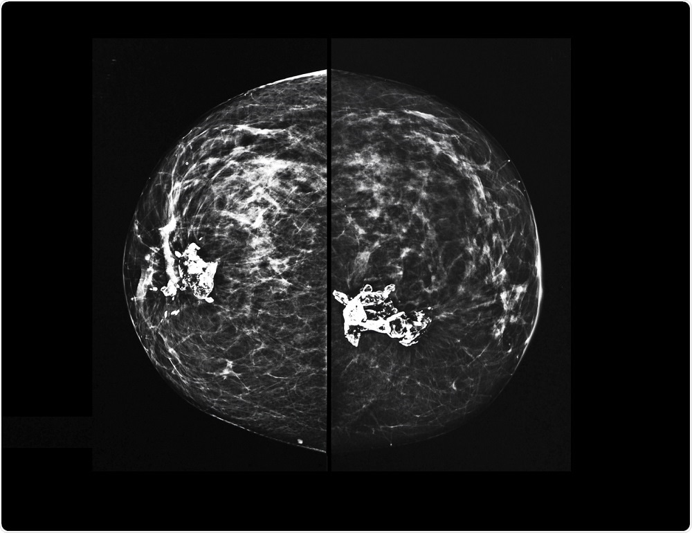
Ductal carcinoma in situ (dcis) is a precancerous state in which cells lining the milk ducts look like cancer cells, but the cells have not spread through the walls of the ducts. Finding breast lumps and seeing change in the size and shape. Generally, whiter mammogram images indicate denser breasts. They're often easy to move around (mobile) and may be tender. The outer edges of these cells look fuzzy or spiky (called spiculated). Doctors use a mammogram to look for early signs of breast cancer. Tumors may be benign or cancerous. In this mammogram image, the breast calcifications are in ductal patterns. The rate of breast cancers discovered as dcis is thought to be increasing, but this is partially a testament to the effectiveness of mammographic breast cancer screening programs.ductal carcinoma in situ represents up to 30% of all new cases of breast cancer discovered by breast cancer screening. Invasive breast cancer can appear as a white patch or mass on a mammogram. However, cancer can also be seen as white, making it a bit obscure in women with extremely dense breasts. It's usually firm and rubbery, and doesn't hurt. Regular mammograms are the best tests doctors have to find breast cancer early, sometimes up to three years before it can be felt.
What does cancer look like on a mammogram? A mammogram can show breast changes such as calcifications, masses, or other symptoms that might be cancer. Abnormalities such as cancerous tumors usually appear brighter because they are denser. Cancers may be seen as masses (like a ball, but usually with an irregular shape), areas of asymmetry that resemble normal tissue, calcifications (white specks), and/or areas of architectural distortion (imagine the puckering caused by pulling a thread in a piece of fabric). This is considered an abnormal mammogram, but not necessarily one that's indicative of cancer.
3d Mammograms Increasing In Popularity For Breast Cancer Screening from www.news-medical.net What does breast cancer look like on a mammogram? Any area that does not look like normal tissue is a possible cause for concern. Any area that does not look like normal tissue is a possible cause for concern. Cancers may be seen as masses (like a ball, but usually with an irregular shape), areas of asymmetry that resemble normal tissue, calcifications (white specks), and/or areas of architectural distortion (imagine the puckering caused by pulling a thread in a piece of fabric). Any area that does not look like normal tissue is a possible cause for concern. If we find these precancerous cells, we can essentially prevent cancer, baker says. A tumor that is benign, it is not a health problem and it may not grow or change shape. Finding breast lumps and seeing change in the size and shape.
Ductal carcinoma in situ (dcis) is a precancerous state in which cells lining the milk ducts look like cancer cells, but the cells have not spread through the walls of the ducts.
Rate of dcis discovery is increasing, due to screening mammograms. How can mammograms be used? Calcifications are calcium deposits within the breast tissue and they look like small white spots. What does breast cancer look like on a mammogram? Cncer a for sure thing? Regular mammograms are the best tests doctors have to find breast cancer early. Abnormalities such as cancerous tumors usually appear brighter because they are denser. Invasive breast cancer can appear as a white patch or mass on a mammogram. The amount of dense breast tissues differs in every woman, and experts usually describe the density levels in grades (1 to 4). There are few risks associated with mammography. Any area that does not look like normal tissue is a possible cause for concern. If we find these precancerous cells, we can essentially prevent cancer, baker says. The rate of breast cancers discovered as dcis is thought to be increasing, but this is partially a testament to the effectiveness of mammographic breast cancer screening programs.ductal carcinoma in situ represents up to 30% of all new cases of breast cancer discovered by breast cancer screening.
Calcifications are tiny flecks of calcium — like grains of salt — in the soft tissue of the breast that can sometimes indicate the presence of an early breast cancer. Doctors use a mammogram to look for early signs of breast cancer. This is considered an abnormal mammogram, but not necessarily one that's indicative of cancer. Cncer a for sure thing? Healthy mammograms can still vary in appearance.
Imaging Inflammatory Breast Cancer Sciencedirect from ars.els-cdn.com Tumors may be benign or cancerous. Breast cancer and some noncancerous (benign) breast conditions can appear white on a mammogram. The tumor cells don't stay within the clear borders of the mass, but instead invade the nearby breast tissue. The look of breast cancer on a mammogram a tumor or lump will appear as a focused white area on the mammogram. The outer edges of these cells look fuzzy or spiky (called spiculated). Cancers may be seen as masses (like a ball, but usually with an irregular shape), areas of asymmetry that can resemble normal tissue, calcifications (white specks), and/or areas of architectural distortion (imagine the puckering caused by pulling a thread in a piece of fabric). Sometimes it can be difficult to tell the difference between mastitis and inflammatory breast cancer. Calcifications are calcium deposits within the breast tissue and they look like small white spots.
Any area that does not look like normal tissue is a possible cause for concern.
Cancers may be seen as masses (like a ball, but usually with an irregular shape), areas of asymmetry that resemble normal tissue, calcifications (white specks), and/or areas of architectural distortion (imagine the puckering caused by pulling a thread in a piece of fabric). Mammograms may show suspicious areas of the breast, white spots, and microcalcifications. Calcifications are tiny flecks of calcium — like grains of salt — in the soft tissue of the breast that can sometimes indicate the presence of an early breast cancer. Doctors use a mammogram to look for early signs of breast cancer. Abnormalities such as cancerous tumors usually appear brighter because they are denser. If you get an abnormal result for mammography and you have had uterine cncer and just checked your cancer antigen its 6.3. What does a suspicious area look like on a mammogram? They're often easy to move around (mobile) and may be tender. Regular mammograms are the best tests doctors have to find breast cancer early, sometimes up to three years before it can be felt. Invasive breast cancer can appear as a white patch or mass on a mammogram. A lump or tumor will show up as a focused white area on a mammogram. What does breast cancer look like on a mammogram? There are few risks associated with mammography.